Skin delivery in a nutshell
Physiology background
To understand skin drug delivery, first we need to get some knowledge about the skin itself. A basic principle in physiology, the branch of biology that studies the normal functions of living organisms and their parts, is that form and function are interconnected. That means, the form and characteristics of a certain cell, tissue or organ have the purpose of allowing the main function of that organ to be successfully performed. For instance, a muscle’s anatomy is evolved to be able to produce movement under the right conditions. Having this concept in mind helps to understand how the skin’s structure is. The skin is the largest organ in the human body and the first barrier against the outside world. Accordingly, this special organ developed all the characteristics necessary to successfully filter what goes in and out of our organism, hence protecting us against different external harms, including pathogens.
Basic skin histology and anatomy
Overview of Skin Structure
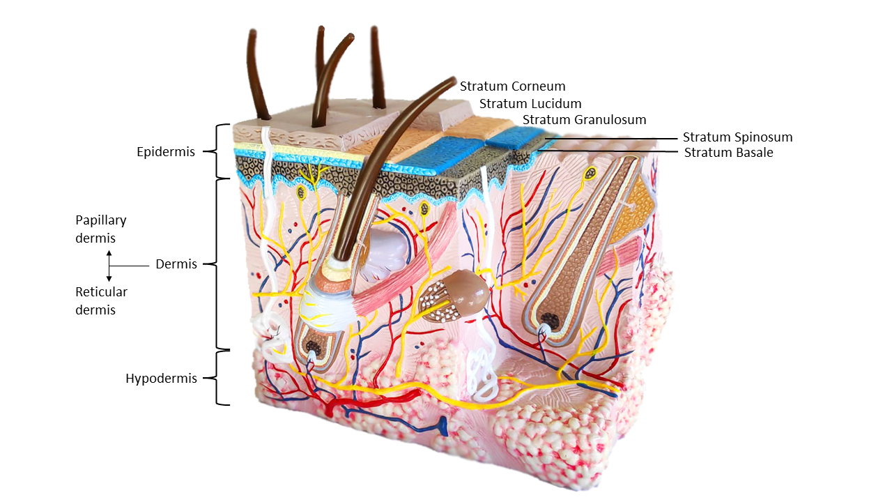
The skin is a multifunctional organ part of the integumentary system with unique anatomy and cellular composition. It consists of layers functioning in a dynamic and interconnected way [2], see Figure 1:
The Epidermis
The epidermis, the outermost layer of the skin and therefore the layer in direct contact with the environment. Consequently, this layer behaves as a physical barrier and is not vascularized to avoid systemic entrance of undesired hazards. The epidermis is formed primarily by keratinocytes, a special cell type able to produce keratin that provides strength. This skin layer is divided in stratums and the outermost one, called stratum corneum is composed of a special cell type called corneocytes, interspersed in lipids, forming the so-called “brick and mortar model”. These are dead cells full of keratin which pileup over each other functioning as a “wall”. It also contains immune cells in case a pathogen breaches the barrier. Additionally, this layer hosts beneficial microbes that contribute to the skin’s protection against environmental threats.
The Dermis
Beneath the epidermis lies the dermis, mainly composed of connective tissue and characterized by abundant extracellular matrix (ECM), which supports the epidermis in nutrition and other aspects. That means, differently from the epidermis this layer is vascularized, therefore it could potentially represent an easy entrance door for microorganisms. That’s why it contains several types of immune cells in great numbers that will “guard” the location, identifying possible “invasions”. These immune cells are mainly located in the papillary dermis which is the upper region of the dermis.
The Hypodermis
The hypodermis which is right under the dermis. This is also known as the subcutaneous tissue, which contains a lot of fat and connective tissue. It supports and drains the dermis through its large lymphatic vessels and veins.
Skin Appendages
Each layer has a different organization and composition that functions as a role to defend us from environmental hazards. Moreover, the skin contains appendages such as hair follicles, sweat and sebaceous glands which are important for accomplishing the many skin functions.